What Is VTach? Understanding the V Tach ECG
Ventricular Tachycardia (VTach) is a potentially life-threatening arrhythmia where the heart’s ventricles beat rapidly and out of sync with the atria. In a VTach ECG, this presents as a wide-complex tachycardia, typically ranging from 150 to 250 beats per minute.
This rapid rhythm prevents the ventricles from filling properly, reducing cardiac output and oxygen delivery to the body. VTach can be monomorphic (consistent QRS shape) or polymorphic (varied QRS shapes), and it may start suddenly without warning.
VTach ECG Characteristics:
- Wide QRS complexes (>0.14 seconds)
- No preceding P waves
- Regular rhythm (though may be slightly irregular)
- No discernible atrial activity
Symptoms of VTach:
- Rapid heartbeat
- Lightheadedness or fainting
- Chest pain or palpitations
- Shortness of breath
Sustained VTach lasts longer than 30 seconds and requires immediate medical attention. If the patient is pulseless, it must be treated like VFib—with immediate defibrillation.
VTach vs VFib ECG: Spot the Difference
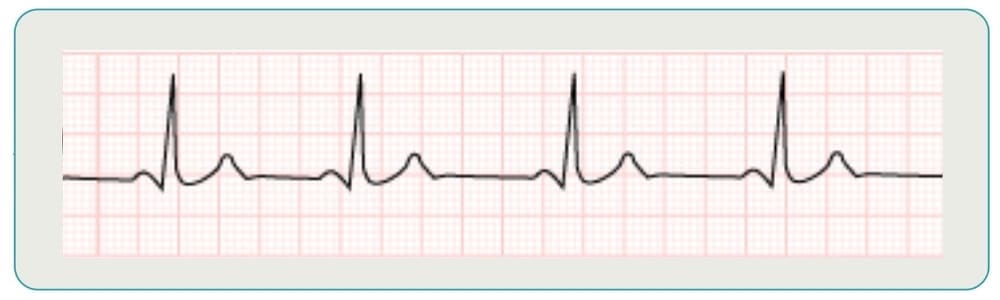
The first ECG is a normal sinus rhythm, with an organized pattern. P waves, QRS complexes, and T waves show up as 3 distinct waves, equally spaced and similar in size.
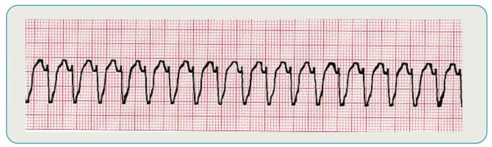
The second ECG shows Vtach, with wide, irregular QRS complexes and occasional P waves. It is difficult to see separation between the QRS complex and the T wave.
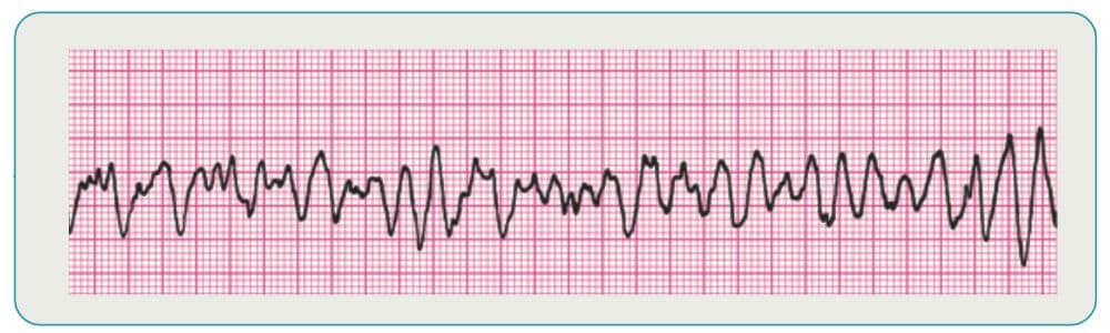
The final ECG shows Vfib, with no clear P waves, T waves, or QRS complexes. The rate may be as fast as 600 beats per minute and the deflections will vary and often decrease as time goes on.
What Is VFib? Interpreting a V-Fib Rhythm Strip
Ventricular Fibrillation (VFib) is a chaotic, disorganized electrical activity in the heart’s ventricles, leading to ineffective pumping and sudden cardiac arrest. A v-fib rhythm strip or ventricular fibrillation EKG shows no identifiable waveforms, just erratic fibrillatory waves.
This arrhythmia is the most common cause of sudden cardiac death. The ventricles quiver instead of contract, and as a result, no blood is pumped from the heart. Immediate recognition and treatment are essential.
VFib ECG Characteristics:
- No clear QRS, P, or T waves
- Disorganized, rapid rhythm (up to 600 bpm)
- Coarse VFib: waves >3mm
- Fine VFib: waves <3mm, resembling a near-flatline
Symptoms Prior to VFib:
- Sudden collapse
- Loss of consciousness
- Gasps or abnormal breathing
Immediate CPR and defibrillation are critical to survival.
VTach vs VFib on ECG: Key Differences Explained
VTach and VFib can look similar at a glance, especially to the untrained eye. But their differences are crucial for treatment decisions. VTach is more organized, with identifiable QRS complexes, while VFib is entirely chaotic with no structure.
Feature | VTach (v tach ECG) | VFib (ventricular fibrillation EKG) |
QRS Complex | Wide and regular | None discernible |
Rhythm | Rapid and regular | Chaotic and disorganized |
Heart Rate | 150–250 bpm | Up to 600 bpm |
P Waves | Absent or dissociated | Absent |
Risk | Can lead to VFib | Immediate cardiac arrest |
Causes, Diagnosis, and Risk Factors
Both VTach and VFib are associated with underlying heart disease or electrical abnormalities. VTach is often linked to scar tissue from a previous heart attack, while VFib frequently follows acute myocardial infarction or severe ischemia.
Common Causes:
- Coronary artery disease
- Prior myocardial infarction
- Cardiomyopathy
- Electrolyte imbalances (e.g., low potassium or magnesium)
- Drug toxicity (e.g., digoxin, antiarrhythmics)
- Congenital heart syndromes (e.g., Long QT, Brugada)
Diagnosis:
- 12-lead ECG during symptoms
- Continuous telemetry or Holter monitoring
- Cardiac event recorders for intermittent episodes
Early diagnosis and intervention reduce the risk of sudden cardiac death.
Treatment for VTach and VFib: What to Do in an Emergency
Early intervention saves lives. The treatment depends on whether the patient is stable and whether a pulse is present.
VTach Treatment:
- Stable: IV antiarrhythmics (amiodarone, lidocaine)
- Unstable: Electrical cardioversion
- Pulseless: Immediate CPR and defibrillation
VFib Treatment:
- Immediate CPR
- Early defibrillation (AED use in the field)
- Advanced cardiac life support (ACLS) protocol with epinephrine, amiodarone
After resuscitation, patients may receive an implantable cardioverter-defibrillator (ICD) or undergo catheter ablation to prevent recurrence.
How to Read ECG Results Like a Pro
Mastering ECG interpretation requires a systematic approach. Focus on each part of the waveform to identify potential abnormalities.
- P-Waves: Look for presence, consistency, and relation to QRS
- PR Interval: Normal is 0.12–0.20 seconds; prolongation may indicate AV block
- QRS Complex: Duration and morphology; wide complexes suggest ventricular origin
- Heart Rate: Use large box method (300 / number of large boxes between R waves)
- Overall Rhythm: Regular vs irregular; evaluate atrial and ventricular activity
Pro Tip: Use the ADD Rule
- Amplitude: Height of each wave reflects voltage
- Deflection: Direction the wave moves (up/down from baseline)
- Duration: Width of waveforms and intervals; longer durations may indicate conduction delays
Why EKG Certification Matters for Healthcare Providers
Quickly recognizing v-tach vs v fib can mean the difference between life and death. In emergency settings, time is critical. Certified providers are trained to:
- Identify dangerous arrhythmias with confidence
- Initiate immediate and appropriate treatment
- Work effectively within code teams
Certification also ensures compliance with workplace and hospital policies and strengthens your credentials for career advancement.
EKG Training with SureFire CPR
At SureFire CPR, our EKG Certification Course offers comprehensive training designed for healthcare professionals. Whether you’re new to ECG interpretation or need a refresher, we provide:
- Real-world ECG interpretation scenarios
- Expert instruction by seasoned professionals
- Same-day online completion certificate
- Up to 10 hours of CE credits available
You’ll leave our course ready to recognize arrhythmias like v tach ECG and v fib rhythm strip—and respond with confidence.
Explore More:
- BLS Certification Course
- ACLS Certification Course
- PALS Certification Course
Final Takeaway: Recognize and Respond to Dangerous Arrhythmias
Understanding how to read v tach ecg and interpret a v fib rhythm strip is crucial for anyone in emergency medicine. These arrhythmias demand rapid recognition and decisive action.
With SureFire CPR’s expert training, you’ll build the confidence to act fast when every second counts. Whether you’re a nurse, paramedic, or medical student, our EKG course gives you the tools to save lives.
Ready to improve your rhythm recognition? Check our EKG class schedule and get certified today.
FAQs: VTach, VFib, and ECG Interpretation
What does a v tach ECG look like?
A: Wide, fast QRS complexes with no P waves and a regular rhythm.
What is a v fib rhythm strip?
A: A disorganized, chaotic waveform with no identifiable QRS, P, or T waves.
Can VTach turn into VFib?
A: Yes. Sustained VTach can degrade into VFib, increasing risk of sudden cardiac arrest.
How fast is the heart rate in VFib?
A: It can reach up to 600 bpm, though no organized beats are present.
Can these arrhythmias be prevented?
A: In some cases, yes. Managing risk factors and using medications or ICDs can help prevent recurrence.